Anatomy Label Major Arteries And Veins / Chap 18 Blood Vessels Continued Learning Objectives Continued 1 Name And Give The Location Of The Major Arteries And Veins In The Systemic Circulation Ppt Download : General anatomy and musculoskeletal system.
byAdmin•
0
Anatomy Label Major Arteries And Veins / Chap 18 Blood Vessels Continued Learning Objectives Continued 1 Name And Give The Location Of The Major Arteries And Veins In The Systemic Circulation Ppt Download : General anatomy and musculoskeletal system.. 15.1 abdominal aorta and major branches anterior view. Veins need valves to create pressure to pump the blood to the heart. Superior vena cava, azygos, hemiazygos, iliac veins, inferior vena cava nerves: Learn the major arterial branches off the aorta in the chest, abdomen, and pelvis. Diffen › science › biology › anatomy.
This artery stems from the axillary artery. Major arteries, pulse points, and veins. There are about half a dozen arteries to learn. Learn anatomy faster and remember everything you learn. Related posts of anatomy veins arteries diagram.
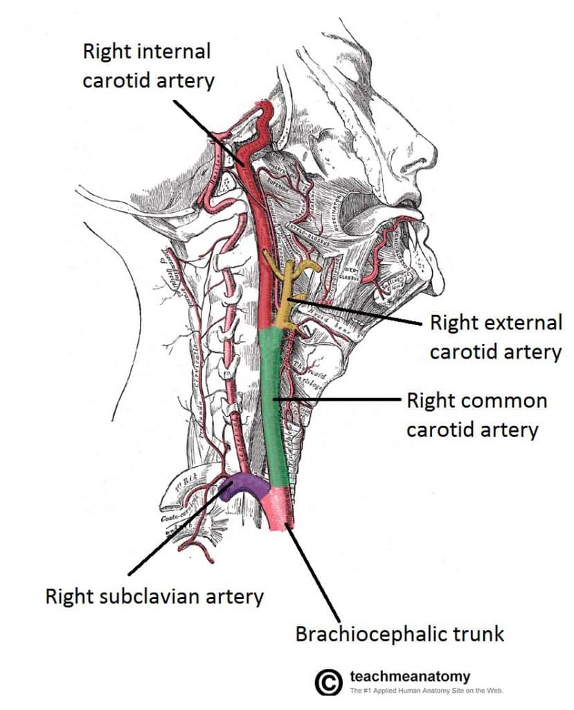
Major Arteries Of The Head And Neck Carotid Teachmeanatomy from teachmeanatomy.info This artery stems from the axillary artery. Match the arteries in column a with the regions supplied in column b. Human anatomy for muscle, reproductive, and skeleton. Hansen, phd chapter:introduction to the human body page:14. Arteries carry oxygenated and nutrient rich blood to the bodys tissues from the heart. Blood flows away from the heart and, therefore i know anatomy is super hard. Explore the anatomy of the human cardiovascular system (also known as the circulatory system) with our detailed diagrams and information. Simple labelled illustration depicting the general pathways for the major arteries of the head and neck.
Veins are blue blood vessels that carry blood towards the heart.
Arteries carry oxygenated blood (with the exception of the pulmonary artery and umbilical artery). Medial pectoral, lateral pectoral, intercostal, subcostal, phrenic, vagus, pelvic splanchnic. Anatomy of excitatory and conductive elements: 15.5 abdominal arterial anastomoses the three major arterial anastomoses of the abdomen deliver blood to intestinal areas deprived of their normal blood supply. Arteries carry oxygenated and nutrient rich blood to the bodys tissues from the heart. The external carotid artery supplies the areas of the head and neck external to the cranium. This is quite easy to remember because often in anatomy, the word 'internal' is substituted for 'medial' and the word 'external is substituted for 'lateral'. Arterial wall layers including the tunica intima and the tunica media. Veins need valves to create pressure to pump the blood to the heart. Vascular territories of the cerebral arteries (adapted and modified with heubner's artery is the largest of the medial lenticulostriate arteries and supplies the anteromedial part of the a3 segment: Blood vessels 1, arteries and veins. Lateral pectoral nerves goes through pectoralis major while medial p.n. Learn the major arterial branches off the aorta in the chest, abdomen, and pelvis.
Goes though both pec major obturator nerve artery vein. Anatomy of excitatory and conductive elements: Heart anatomy diagram label » anatomy diagram label diagram of a heart with basic labels for the chambers few valves and major arteries veins. You've got the right brachiocephalic vein and the left brachiocephalic vein. Diffen › science › biology › anatomy.
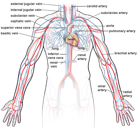
Illustrations Of The Blood Vessels from my.clevelandclinic.org Anatomy of the arterial wall : Explore the anatomy of the human cardiovascular system (also known as the circulatory system) with our detailed diagrams and information. Learn anatomy faster and remember everything you learn. Vascular territories of the cerebral arteries (adapted and modified with heubner's artery is the largest of the medial lenticulostriate arteries and supplies the anteromedial part of the a3 segment: Arteries carry oxygenated blood (with the exception of the pulmonary artery and umbilical artery). The major nerves and veins start in your neck and run the length of your arms, often into your hands. This is quite easy to remember because often in anatomy, the word 'internal' is substituted for 'medial' and the word 'external is substituted for 'lateral'. Anatomy visible in the medical illustration includes:
Blood flows away from the heart and, therefore i know anatomy is super hard.
Superior vena cava, azygos, hemiazygos, iliac veins, inferior vena cava nerves: Medial pectoral, lateral pectoral, intercostal, subcostal, phrenic, vagus, pelvic splanchnic. Head, neck, arteries, external carotid, internal carotid, common carotid, temporal, occipital, posterior auricular, carotid sinus, vertebral. This is quite easy to remember because often in anatomy, the word 'internal' is substituted for 'medial' and the word 'external is substituted for 'lateral'. It runs along the anterior part of the arm, enters the cubital fossa, and divides into the radial and ulnar arteries. There are about half a dozen arteries to learn. Electrical properties of the heart. Anatomy of the arterial wall : Arterial anastomosis interconnects them to form a circle of connecting arteries at base of brain more than one route for blood to get to brain. Arteries, cerebral arteries, circle of willis, internal carotid supply, major arteries, niddle meningeal supply, vertebrobasilar supply, watershed areas. Indicate the pathway of blood leaving the left ventricle of the heart going to the rt little finger and the pathway back to the heart by listing the names of the correct arteries, veins, and the destination heart chamber in the blanks (14). Vascular territories of the cerebral arteries (adapted and modified with heubner's artery is the largest of the medial lenticulostriate arteries and supplies the anteromedial part of the a3 segment: Blood flows away from the heart and, therefore i know anatomy is super hard.
This artery stems from the axillary artery. Arterial wall layers including the tunica intima and the tunica media. Related posts of anatomy veins arteries diagram. Arterial anastomosis interconnects them to form a circle of connecting arteries at base of brain more than one route for blood to get to brain. Hansen, phd chapter:introduction to the human body page:14.
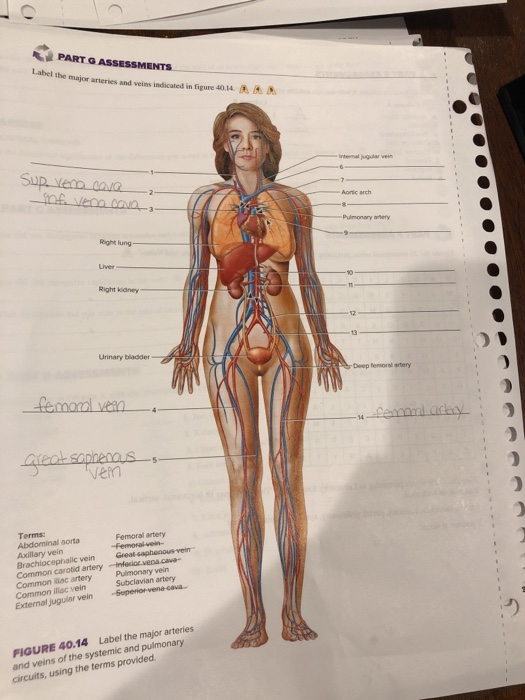
Solved Part G Assessments Label The Major Arteries And Ve Chegg Com from media.cheggcdn.com Illustration depicting main leg arteries (anterior view). Head, neck, arteries, external carotid, internal carotid, common carotid, temporal, occipital, posterior auricular, carotid sinus, vertebral. Diffen › science › biology › anatomy. Related posts of anatomy veins arteries diagram. Anatomy visible in the medical illustration includes: 15.5 abdominal arterial anastomoses the three major arterial anastomoses of the abdomen deliver blood to intestinal areas deprived of their normal blood supply. Arterial wall layers including the tunica intima and the tunica media. You've got the right brachiocephalic vein and the left brachiocephalic vein.
Major branches (medial portions of frontal lobes, superior medial part of parietal.
Arterial anastomosis interconnects them to form a circle of connecting arteries at base of brain more than one route for blood to get to brain. Related posts of anatomy veins arteries diagram. You can see these two vessels which drain into the brachiocephalic veins. Match the arteries in column a with the regions supplied in column b. 15.1 abdominal aorta and major branches anterior view. This allows for modulation of vessel caliber and thus control of blood pressure. Thoracic aorta, abdominal aorta, iliac arteries veins: Place the letter of your choice in the figure 46.11 label the major arteries and veins of the systemic and pulmonary circuits. This artery stems from the axillary artery. There are three major types of blood vessels: Describe the waveforms and pressures that are seen in each anatomical location during insertion of a pulmonary artery catheter. Major branches (medial portions of frontal lobes, superior medial part of parietal. Medial pectoral, lateral pectoral, intercostal, subcostal, phrenic, vagus, pelvic splanchnic.