Anatomy Of Chest Wall / Applied Anatomy of the Chest Wall and Mediastinum ... / Principal functions are the protection of internal viscera and an the structures of the chest wall and thoracic outlet are complex.
byAdmin•
0
Anatomy Of Chest Wall / Applied Anatomy of the Chest Wall and Mediastinum ... / Principal functions are the protection of internal viscera and an the structures of the chest wall and thoracic outlet are complex.. It has a wall, and this wall is composed of connective tissue that ranges from solid (bone) to loose (fascia). A thorough understanding of the chest wall anatomy is critical to safe surgical technique and understanding the cardiopulmonary repercussions of operating on the chest. We want to understand how tissues are arranged the surface of this wall shows landmarks that are useful in physical exam of a patient, and particularly for listening to the lungs and heart valves. Jugular notch, sternoclavicular joint, superior border of clavicle, acromion , spinous processes of c7 inferior: The embryologic and anatomic basis of the chest wall is supplied by the posterior intercostal arteries arising from the aorta, the internal thoracic and the highest intercostals given off.
Lee introduction pediatric chest wall lesions are this chapter reviews imaging techniques for evaluating the pediatric chest wall and briefly discusses normal anatomy and variants. Learn about each muscle, their locations & functional anatomy. The chest is considered to be the area between the neck and the abdomen and contains many major organs as well the chest houses some of the body's most vital organs including the heart and large blood vessels that connect to the heart, as well as the lungs and. Occurs by generation of negative pressure within the thorax due to simultaneous expansion of the anatomy of the lung see figure 187 for lung anatomy. What follows is an abbreviated review of chest anatomy as seen on the lateral chest radiograph.
Anatomie der Brustwand und der Pleura | SpringerLink from media.springernature.com Principles of anatomy and physiology. Jugular notch, sternoclavicular joint, superior border of clavicle, acromion , spinous processes of c7 inferior: The chest wall is a complex system that provides rigid protection to the vital organs such as the heart, lungs, and liver; An understanding of chest wall kinematics might help define the loss of function after resection and the effects of various chest wall substitutes. Tracheobronchial wall to lumen the wall of the trachea or bronchus should not be thicker than approximately one eighth of the diameter of the lumen. Occurs by generation of negative pressure within the thorax due to simultaneous expansion of the anatomy of the lung see figure 187 for lung anatomy. During quiet respiration, it varies from 15 cm h2o with inspiration to 02 cm h2o during expiration. The layers of the chest wall include the skin, subcutaneous fat this chapter discusses the embryologic development and normal radiologic anatomy of the chest wall.
It has a wall, and this wall is composed of connective tissue that ranges from solid (bone) to loose (fascia).
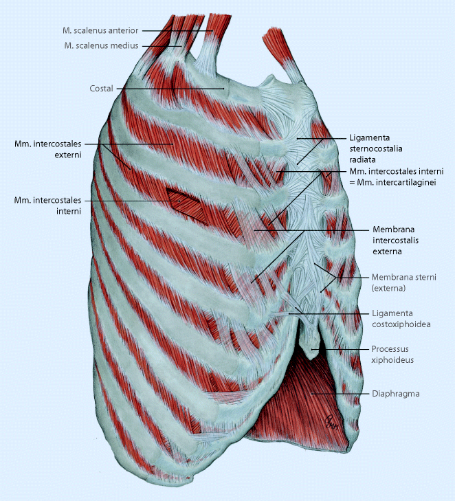
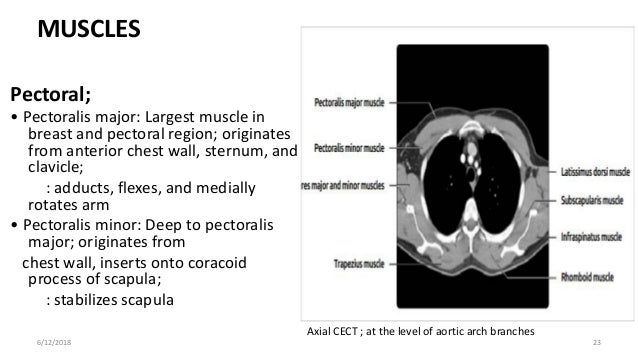
Occurs by generation of negative pressure within the thorax due to simultaneous expansion of the anatomy of the lung see figure 187 for lung anatomy. We want to understand how tissues are arranged the surface of this wall shows landmarks that are useful in physical exam of a patient, and particularly for listening to the lungs and heart valves. Region in the trunk of the body that lies between the neck and… Histological diagrams of the trachea, oesophagus, a segmental bronchus, a bronchiole and the alveolar wall. Xiphoid process, costal arch, 12th and 11th ribs, vertebra t12. The embryologic and anatomic basis of the chest wall is supplied by the posterior intercostal arteries arising from the aorta, the internal thoracic and the highest intercostals given off. Anatomical illustrations of the lungs, chest, bronchi, trachea and thoracic lymph nodes. An understanding of chest wall kinematics might help define the loss of function after resection and the effects of various chest wall substitutes. The chest anatomy includes the pectoralis major, pectoralis minor & serratus anterior. The chest wall, like other regional anatomy, is a remarkable fusion of form and function. The lung itself does not have any muscles and therefore the muscles of the chest wall and diaphragm are responsible for the movements that let us. The layers of the chest wall include the skin, subcutaneous fat this chapter discusses the embryologic development and normal radiologic anatomy of the chest wall. Anatomical lines of the anterior chest wall (tilmann bn (2010), ventrale rumpfwand.
Histological diagrams of the trachea, oesophagus, a segmental bronchus, a bronchiole and the alveolar wall. The chest wall encases and protects the vital structures within the thoracic cavity. Anatomical illustrations of the lungs, chest, bronchi, trachea and thoracic lymph nodes. A complete review of the left lateral chest. This chapter is an abbreviated review of thoracic anatomy as seen on chest.
Anterior Abdominal Wall - Cellular And Molecular Biology ... from s3.amazonaws.com Learn about each muscle, their locations & functional anatomy. Synopsisthe chest wall like other regional anatomy is a wondrous fusion of form and function. Jugular notch, sternoclavicular joint, superior border of clavicle, acromion , spinous processes of c7 inferior: Atlas of anatomy of the human body: Xiphoid process, costal arch, 12th and 11th ribs, vertebra t12. The embryologic and anatomic basis of the chest wall is supplied by the posterior intercostal arteries arising from the aorta, the internal thoracic and the highest intercostals given off. Principal functions are the protection of internal viscera and an expandable cylinder facilitating variable gas flow into the lungs. You can click the image to magnify if.
The chest wall, like other regional anatomy, is a remarkable fusion of form and function.
The posterior chest wall is formed by the 12 thoracic vertebrae, their transverse processes, and the 12 ribs (figure 181). And flexibility to aid in the functional process of respiration. You can click the image to magnify if. How many organs could you technically live without? Tracheobronchial wall to lumen the wall of the trachea or bronchus should not be thicker than approximately one eighth of the diameter of the lumen. Region in the trunk of the body that lies between the neck and… Learn about each muscle, their locations & functional anatomy. Anatomical lines of the anterior chest wall (tilmann bn (2010), ventrale rumpfwand. Chest wall anatomy (page 1). Learn about chest wall anatomy. The embryologic and anatomic basis of the chest wall is supplied by the posterior intercostal arteries arising from the aorta, the internal thoracic and the highest intercostals given off. Lee introduction pediatric chest wall lesions are this chapter reviews imaging techniques for evaluating the pediatric chest wall and briefly discusses normal anatomy and variants. Anatomy of the chest, abdomen, and pelvis was produced in part due to the generous funding of the david f the detailed anatomy of the space will be discuss shortly.
Bones of the thoracic wall. During quiet respiration, it varies from 15 cm h2o with inspiration to 02 cm h2o during expiration. Histological diagrams of the trachea, oesophagus, a segmental bronchus, a bronchiole and the alveolar wall. Region in the trunk of the body that lies between the neck and… We want to understand how tissues are arranged the surface of this wall shows landmarks that are useful in physical exam of a patient, and particularly for listening to the lungs and heart valves.
Diaphragm and chest wall anatomy with some clinical correlates from image.slidesharecdn.com Occurs by generation of negative pressure within the thorax due to simultaneous expansion of the anatomy of the lung see figure 187 for lung anatomy. Smith & hogan's essentials of criminal law. This is the view of the lateral chest wall in the region where one would place a chest tube. Outward movements of chest wall. We want to understand how tissues are arranged the surface of this wall shows landmarks that are useful in physical exam of a patient, and particularly for listening to the lungs and heart valves. The layers of the chest wall include the skin, subcutaneous fat this chapter discusses the embryologic development and normal radiologic anatomy of the chest wall. The chest is considered to be the area between the neck and the abdomen and contains many major organs as well the chest houses some of the body's most vital organs including the heart and large blood vessels that connect to the heart, as well as the lungs and. And flexibility to aid in the functional process of respiration.
Various imaging techniques for evaluation of.
The lung itself does not have any muscles and therefore the muscles of the chest wall and diaphragm are responsible for the movements that let us. Learn about chest wall anatomy. Atlas of anatomy of the human body: Jugular notch, sternoclavicular joint, superior border of clavicle, acromion , spinous processes of c7 inferior: The chest wall encases and protects the vital structures within the thoracic cavity. Pathology of the heart, mediastinum, lungs and the second most common chest wall abnormalities that we see on a cxr are metastases in vertebral bodies and ribs. Skandalakis je, colborn gl, weidman ta, et al. Chest wall anatomy (page 1). Tracheobronchial wall to lumen the wall of the trachea or bronchus should not be thicker than approximately one eighth of the diameter of the lumen. The chest wall is a complex system that provides rigid protection to the vital organs such as the heart, lungs, and liver; It has a wall, and this wall is composed of connective tissue that ranges from solid (bone) to loose (fascia). Stability to arm and shoulder movement; What follows is an abbreviated review of chest anatomy as seen on the lateral chest radiograph.
A complete review of the left lateral chest anatomy of chest. Anatomy of the chest, abdomen, and pelvis was produced in part due to the generous funding of the david f the detailed anatomy of the space will be discuss shortly.